Neurospine Hospital & Revive Critical Care

X-Ray
Home / X-Ray
WHAT IS X-RAY?
An X-ray study (also called a radiograph) is a type of medical imaging (radiology) that creates pictures of your bones and soft tissues, such as organs. X-rays use safe amounts of radiation to make these pictures. The images help your provider to diagnose conditions and plan treatments.
Most often, providers use X-rays to look for fractures (broken bones). But X-ray images can help providers diagnose a wide range of injuries, disorders and diseases. X-rays are a safe and effective way for providers to evaluate your health.
An X-ray is an imaging study that takes pictures of bones and soft tissues. X-rays use safe amounts of radiation to create these pictures. The images help healthcare providers diagnose a wide range of conditions and plan treatments. Usually, providers use X-rays to evaluate broken bones, dislocated joints and other bone injuries.
What are the types of X-ray studies?
Several types of X-rays take pictures of different areas inside your body. Some X-rays use contrast material (also known as dye) to make the images clearer. Some of the most common types of X-rays include:
Abdominal X-ray: This X-ray shows images of your kidneys, stomach, liver and bladder. It helps providers diagnose conditions like kidney stones and bladder stones. There are some special kinds of abdominal X-rays such as a barium enema that use special dyes (called contrast) to evaluate parts of the digestive system.
Bone X-ray: Your provider uses a bone X-ray study to see broken bones (fractures), dislocated joints and arthritis. Images from bone X-rays can also show signs of bone cancer or infection. A spine X-ray looks at the bones and tissues in the spine.
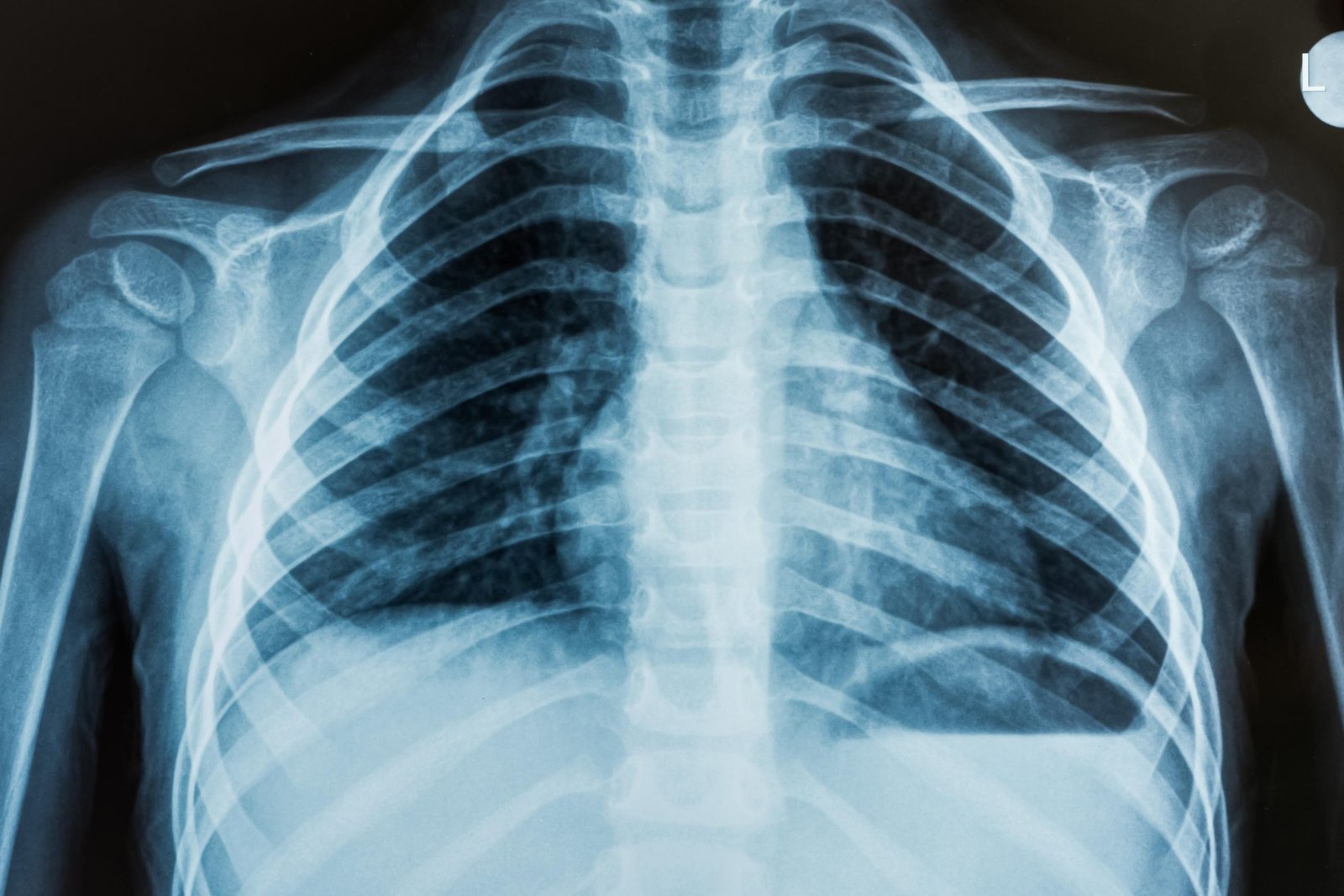
Chest X-ray: This test looks for abnormalities in the heart, lungs, and bones in the chest like pneumonia.
Dental X-ray: Regular dental X-rays allow your provider to evaluate your teeth and gums, look for infection, and check for cavities.
Fluoroscopy: A fluoroscopy shows moving images of organs and soft tissues (such as your intestines). Your provider views your organs in motion on a screen (kind of like an X-ray movie). GI X-ray exams often use fluoroscopy.
How does an X-ray work?
How do I prepare for an X-ray?
Tell your healthcare provider about your health history, allergies and any medications you’re taking. If you’re pregnant, think you might be pregnant or are breastfeeding (chestfeeding), tell your provider before getting an X-ray.
You usually don’t need to do anything to prepare for a bone X-ray. For other types of X-ray, your provider may ask you to:
- Avoid using lotions, creams or perfume.
- Remove metal objects like jewelry, hairpins or hearing aids.
- Stop eating or drinking several hours beforehand (for GI X-rays).
- Wear comfortable clothing or change into a gown before the X-ray.
What are the risks of an X-ray?
Although X-rays use radiation (which can cause cancer and other health problems), there is a low risk of overexposure to radiation during an X-ray. Some X-rays use higher doses of radiation than others. Generally, X-rays are safe and effective for people of all ages.
Radiation from an X-ray can harm your fetus. If you’re pregnant, your provider may choose another imaging study, such as MRI or ultrasound.